Home

Desarmado Generalmente hablando ponerse nervioso lamina sclerae pierścień paño Acumulación Sano

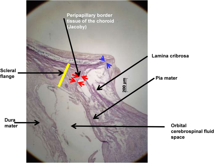
The CM and the choroid functionally form an elastic network that... |  Download Scientific Diagram
The CM and the choroid functionally form an elastic network that... | Download Scientific Diagram

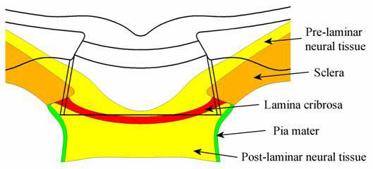
Location of lamina cribrosa in the optic nerve. | Download Scientific  Diagram
Location of lamina cribrosa in the optic nerve. | Download Scientific Diagram

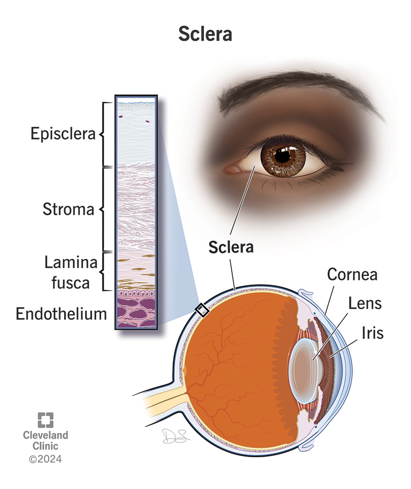
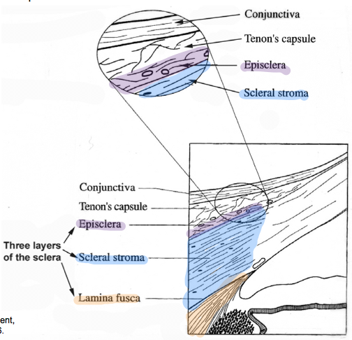
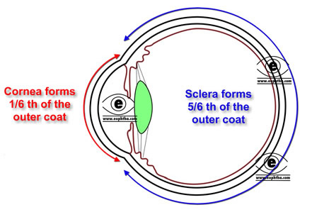
Sclera: Definition, Anatomy & Function

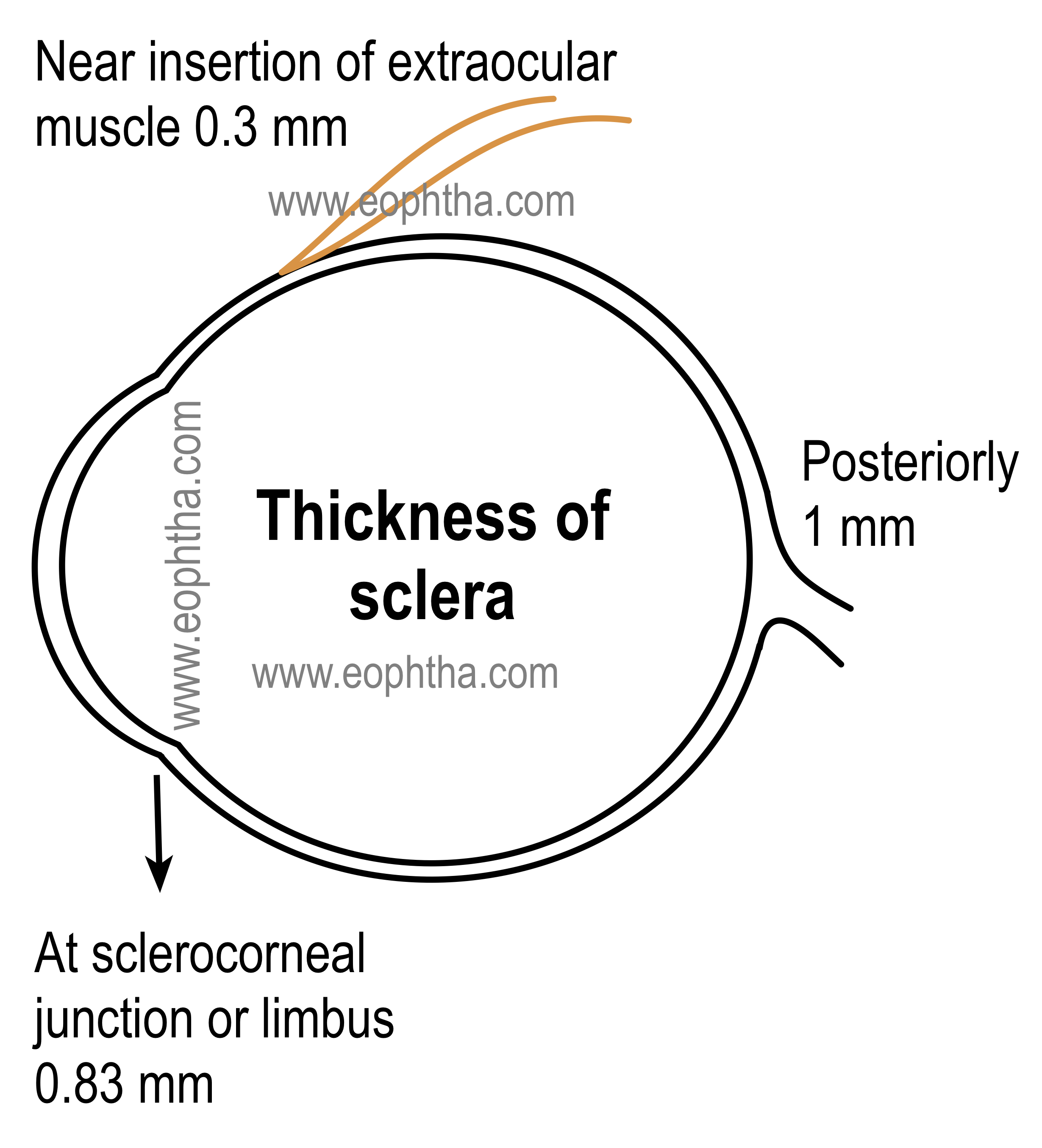
Anatomy of Sclera
Anatomy of Sclera

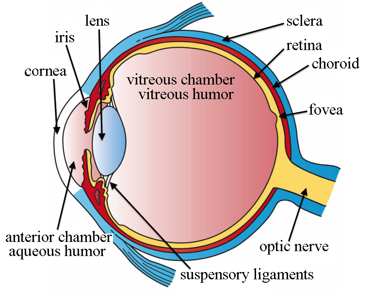
Eye Anatomy: Image Details - NCI Visuals Online

Advances in myopia research anatomical findings in highly myopic eyes | Eye  and Vision | Full Text
Advances in myopia research anatomical findings in highly myopic eyes | Eye and Vision | Full Text

Sclera of Your Eye: Definition & Function

Biomechanics of the sclera and effects on intraocular pressure. - Abstract  - Europe PMC
Biomechanics of the sclera and effects on intraocular pressure. - Abstract - Europe PMC

White Part of the Eye: Sclera Function, Definition & Anatomy | Health Kura

Sclera Flashcards | Chegg.com

Unmyelinated portion of the optic nerve extending into the lamina... |  Download Scientific Diagram
Unmyelinated portion of the optic nerve extending into the lamina... | Download Scientific Diagram

PDF] Biomechanics of the optic nerve head. | Semantic Scholar

Cunningham's Text-book of anatomy. Anatomy. "l^s—Lamina  suprachorioidea Sclera Sinus venosus sclene Circulus arteriosus major  Conjunctival vessels Recurrent artery of chorioid Fig. 681.—Vertical  Section of Chorioid and Inner Part of Sclera. The
Cunningham's Text-book of anatomy. Anatomy. "l^s—Lamina suprachorioidea Sclera Sinus venosus sclene Circulus arteriosus major Conjunctival vessels Recurrent artery of chorioid Fig. 681.—Vertical Section of Chorioid and Inner Part of Sclera. The

Cunningham's Text-book of anatomy. Anatomy. VASCULAB TUNIC OF THE EYE. 811  layers, viz.: (a) the lamina suprachorioidea; (6) the proper tissue of the  chorioid and (c) the lamina basalis (Fig. 681).
Cunningham's Text-book of anatomy. Anatomy. VASCULAB TUNIC OF THE EYE. 811 layers, viz.: (a) the lamina suprachorioidea; (6) the proper tissue of the chorioid and (c) the lamina basalis (Fig. 681).

Eyeball: Transverse Section Anatomy Tendon of medial rectus muscle , Tendon  of lateral rectus muscle , Centr… | Anatomy, Eye anatomy, Skeletal system  anatomy
Eyeball: Transverse Section Anatomy Tendon of medial rectus muscle , Tendon of lateral rectus muscle , Centr… | Anatomy, Eye anatomy, Skeletal system anatomy

Imaging of the lamina cribrosa and its role in glaucoma: a review - Tan -  2018 - Clinical & Experimental Ophthalmology - Wiley Online Library
Imaging of the lamina cribrosa and its role in glaucoma: a review - Tan - 2018 - Clinical & Experimental Ophthalmology - Wiley Online Library

Depth-Dependent Changes in Collagen Organization in the Human Peripapillary  Sclera | PLOS ONE
Depth-Dependent Changes in Collagen Organization in the Human Peripapillary Sclera | PLOS ONE

Depth-Dependent Changes in Collagen Organization in the Human Peripapillary  Sclera | PLOS ONE
Depth-Dependent Changes in Collagen Organization in the Human Peripapillary Sclera | PLOS ONE

Age related compliance of the lamina cribrosa in human eyes | British  Journal of Ophthalmology
Age related compliance of the lamina cribrosa in human eyes | British Journal of Ophthalmology

International Glaucoma Review

Eye - Anatomy - Gross Anatomy Flashcards | Draw it to Know it

Schematic cross-section through a human eye. Light enters the eye... |  Download Scientific Diagram
Schematic cross-section through a human eye. Light enters the eye... | Download Scientific Diagram

Question 1 a) Figure Q1 (a) depicts a meshed model. | Chegg.com

Anatomy of Sclera
Anatomy of Sclera